|
Cardiovascular Biomechanics and A.I. Laboratory |
|
|
Imperial College London, Department of bioengineering |

|
Placenta
Biomechanics |
|||||||
|
Placenta
is a powerful organ that support the fetus growth during pregnancy. Abnormal
development of umbilical and placenta will have detrimental effect on fetus,
be it in utero or later in life, and it can also have a serious implication
for maternal health. We advocate a biomechanics approach to understanding
placenta health and diseases. We believe that such an approach can lead to
new insights, leading to better detection, diagnosis, and even treatment. Intrauterine
Growth Restriction (IUGR) We
are particularly interested in IUGR, which is a disease of the placenta where
not enough nutrients and oxygen can be transferred from the mother to the
fetus, leading to 5-10x higher mortality rate, and life-long morbidities such
as neuro-maldevelopment, hypertension, diabetes and cardiovascular diseases.
Even in developed world, its prevalence is high, at 3%. Currently, there are
no proven method to prevent or treat IUGR, and detection rate remains poor,
as it is difficult to differentiate between healthy but small babies and
diseased babies. However, successful detection can allow management
strategies such as timing of delivery, which can improve outcome. Mechanical
Properties of Normal and IUGR Placenta Tissue We
performed mechanical testing of post-delivery human placenta samples, to
characterize mechanical properties, and to understand changes during IUGR.
Placenta tissues were found to have substantial viscoelasticity and are thus
sensitive to loading rates during mechanical testing. They are surprisingly
isotropic in stiffness, as shown after testing the same samples in different
directions. IUGR placenta tissues are stiffer than normal ones, but the
differences are only significant at a low compression rate. At the same time,
IUGR placenta tissues have a higher collagen to elastin ratio than normal
ones. Reference: -
Saw SN, Low JYR, Ong MHH, Poh YW, Mattar CNZ, Biswas A, Yap CH.
"Hyperelastic Mechanical Properties of Ex Vivo Normal and Intrauterine
Growth Restricted Placenta." Ann
Biomed Engr. 2018 Jul 1;46(7):1066-77
Placenta
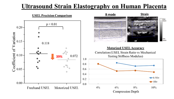
Ultrasound Elastography to Detect IUGR We
investigate whether alternative detection techniques, such as ultrasound
elastography can be successful in detecting IUGR. Informed by our mechanical
testing work, we propose that ultrasound strain elastography should be (1)
performed with a motorized control of the ultrasound transducer (because
viscoelasticity implies that different loading rate can alter tissue
stiffness), (2) measured at a low compression depth and lower compression
rate (our results shows higher correlation between elastography results and
mechanical testing validation), and (3) an external polymeric pad should be
used as the reference layer, instead of bodily tissues, as this ensures that
the reference layers stiffness can be well controlled. Reference: -
Saw SN, Low JYR, Mattar CNZ, Biswas A, Chen L, Yap CH. "Motorizing
and Optimizing Ultrasound Strain Elastography for Detecting Intrauterine
Growth Restriction Pregnancies." Ultrasound
Med. Biol. 2018 Mar 1;44(3):532-43
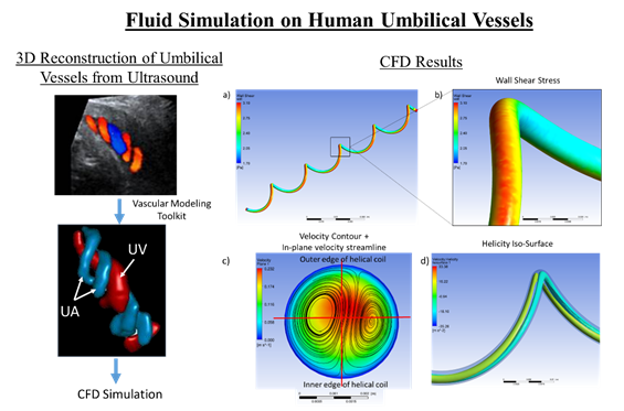
Umbilical
Vascular Fluid Dynamics Using
clinical ultrasound imaging, we extracted anatomy and flow velocities of
umbilical arteries and veins, and performed computational fluid dynamics to
understand relationship between flow forces and vascular sizes. Some
discoveries we made were: (1)
Flow
wall shear stresses in umbilical arteries were independent of size,
suggesting homeostatic mechanisms to maintain certain levels of wall shear
stresses. This was not true for veins. (2)
Flow
profiles departed significantly from a parabolic profile in the arteries, due
to their spiral geometry (3)
Umbilical
arterial flow resistance and wall shear stress environment does not change
substantially during umbilical cord bending, but this is not true for veins (4)
During
IUGR, the wall shear stress environment of arteries and vein did not deviate
from normal pregnancies, suggesting wall shear sensing behaviour may not have
changed. Reference: -
Saw SN, Chia DAK, Biswas A, Mattar CNZ, Yap CH.
"Characterization of the In Vivo Shear Stress Environment of Human Fetus
Umbilical Arteries and Veins." Biomech
Model Mechanobiol. 2017 Feb;16(1):197-211 -
Saw SN, Poh YW, Chia DAK, Biswas A, Mattar CNZ, Yap CH.
"Characterization of the Hemodynamic Wall Shear Stresses in Human
Umbilical Vessels from Normal and Intrauterine Growth Restricted
Pregnancies." Biomech Model
Mechanobiol. 2018 Aug;17(4):1107-1117
Chorionic
Arterial Anatomy, Mechanical Properties and Pulsatility We
performed vascular casting to investigate placenta arterial anatomy,
performed lumped parameter computational modelling to understand pulsatility
in these vessels, and mechanical testing to understand stiffness properties.
Comparisons were made between normal and IUGR human samples. Results
showed that IUGR chorionic arteries were more distensible, and this could
explain the high umbilical pulsatility indices (resistance index, RI, and
pulsatility index, PI). IUGR arteries were smaller than normal ones, but
there were few other differences in terms of vascular and branching geometry,
and opening angle of vessels. Reference: - Saw SN, Tay JJH, Poh YW, Yang L, Tan WC, Tan LK, Clark A, Biswas A, Mattar CNZ, Yap CH. "Altered Placental Chorionic Arterial Biomechanical Properties During Intrauterine Growth Restriction." Scientific Reports. 2018 Nov 8;8(1):16526.
Future
Work Currently,
we are pursuing the use of the rat model of IUGR to understand fetal heart
and vascular growth and remodelling during IUGR, and how the cardiovascular
function, mechanical properties, and structure changes over gestation. |
||||||||
|
|