|
Cardiovascular Biomechanics and A.I. Laboratory |
|
|
Imperial College London, Department of bioengineering |

|
Human
Fetal Heart Echocardiography Image Processing |
|||||||||
|
Motivation Ultrasound
imaging is the de facto modality for fetal heart imaging. It has better
resolution and is more readily available than MRI, and unlike the CT, it does
not use ionizing radiation that can threaten the fetus. However, ultrasound
can be noisy, and is prone to signal losses, and is especially challenging
when applied to structures as small as the human fetal heart. We develop
tools to improve the imaging of the fetal heart, and to extract useful
information on its physiology, function, and biomechanics. |
||||||||||
|
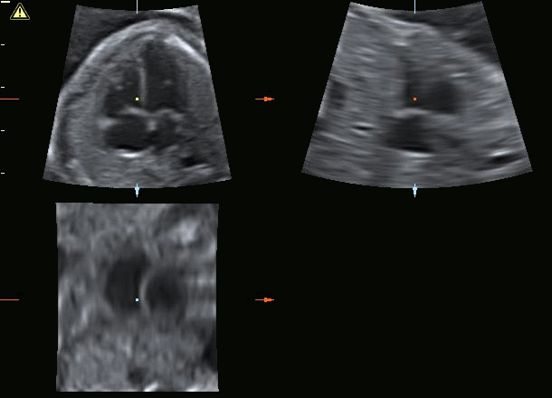
|
Sample of 4D ultrasound images of
a human fetal heart (STIC mode) that is used for computational simulations. |
|||||||||
|
|
Cardiac
Motion Estimation We
developed a method to track the motion of the heart over the cardiac cycle
from fetal echo. 3D free-form deformation pair-wise image registration is
first performed, and a global motion field consisting of spatial B-splines of
temporal Fouriers is then iteratively fitted onto registration velocity field
outputs, to enforce cyclic motions, and temporal and spatial consistency. The
technique is validated using echo data from the cardiac motion analysis
challenge, which comes with MRI truths. Cardiac estimation algorithms often
underestimate the stroke volume. With our approach, additional inputs of
segmentations at when the heart is at its the largest and smallest can be
used to regularize the algorithm to produce motions with accurate stroke
volumes. References:
-
Wiputra H, Chan WX, Foo YY, Ho S, Yap CH. "Cardiac
Motion Estimation from Medical Images: Regularisation Framework Applied on
Pairwise Image Registration Displacement Fields." Scientific
Reports. 2020 Oct 28; 10(1):1-4 |
|||||||||
|
|
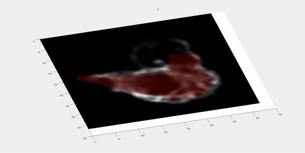
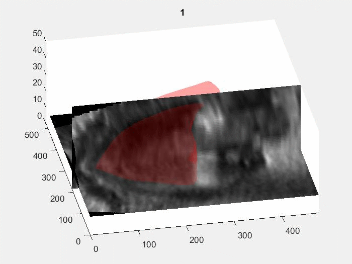
Image registration
tracking luminal space of a 5 days post fertilization of a zebrafish embryo
from 4D microscopy images |
Image registration
tracking of the luminal space of a human fetal right ventricle from 4D
clinical ultrasound images |
||||||||
|
|
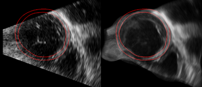
Image
Compounding to Improve Fetal Echocardiography Quality Using
our validated cardiac motion estimation algorithm, we developed an algorithm
for 2D and 3D image compounding of all frames within the cardiac cycle.
Images from all time frames were deformed to match that of the reference
time, and all these images were fused. Thereafter, the fused image can be
re-animated over the cardiac cycle with our cardiac motion model. Compounding
could improve contrast-to-noise ratios, and reveal previously unclear cardiac
structures. Further, if the heart momentarily exceeds the field of view,
compounding enables its recovery, enabling the image to exceed its field of
view.
References:
- Chan WX, Zheng Y, Wiputra H, Leo HL, Yap CH. "Full Cardiac Cycle Asynchronous Temporal Compounding of 3D Echocardiography Images." Med Image Anal. 2021 Dec; 74:102229. Fetal
heart echo scans have low resolution in the lateral and elevational
directions, but high resolution in the axial direction. We thus propose to
scan the heart at various orientations, and perform Multiview compounding
(compounding of scans at various views) to improve resolution and
signals-to-noise.
|
|||||||||
|
|
Future
Work We
seek to continue innovating in image processing techniques, so as to gain better
tools to evaluate fetal heart function, and to detect dysfunction and
congenital malformations. |
|||||||||